
55 In general Candida albicans is the most frequently isolated of the more than 100 known species which include a. The cause or causes of chronic paronychia are not fully understood.

Microbiology Case Study A 65 Year Old Female With Altered Mental Status Lablogatory

Gram Stain Reaction Of Yeast Smear Indicating Gram Positive Yeasts With Download Scientific Diagram
Details Public Health Image Library Phil
The most common causes of.

Candida albicans gram stain. Which of the following microorganisms are not matched correctly with the appropriate isolation media. On Endo agar it looks like lactose negativeAll four strains are mannitol positive best seen in fig. Patients present with vaginal redness itching.
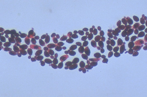
Stains Gram-positive and may be budding or producing pseudohyphae. Gram stain results consistent with a diagnosis of bacterial vaginosis include a markedly reduced or absent lactobacillus morphology. Gram staining is a bacteriological laboratory technique 5 used to differentiate bacterial species into two large groups gram-positive and gram-negative based on.
In urine Candida albicans and other less commonly seen species such as Candida parapsilosis and Candida tropicalis will appear as budding yeasts 410 μm in diameter that often show formation of hyphal elements. D cellobiose negative strains A B. Secondary Candida pneumonia is relatively common but primary Candida pneumonia is rare in other than immunocompromised patients in the intensive care unit.
If you are sick or taking antibiotics it can multiply and cause an infection. Four different strains of Escherichia coli on Endo agar with biochemical slope. The Gram stain is the gold standard according to the CDC and Nugent criterion for reporting the budding yeast cells andor pseudohyphae representative of true vaginal candidiasis.
In addition to the confirmation of candidiasis testing to diagnose an underlying immunocompromising condition is important. Molecular methods may be used to identify Candida species within 60 min from positive blood cultures when the Gram stain is. These microorganisms have great clinical importance in hospitals because they put patients in the intensive care unit ICU at high risk and lead to high morbidity and mortality12 Two large groups Enterobacteriaceae and the non-fermenters are.
Other less common pathogens include Candida albicans Nocardia asteroids Aspergillus HSV. Candida is the scientific name for yeast. Haemorrhage oedema and necrosis are seen in and around the involved vessels.
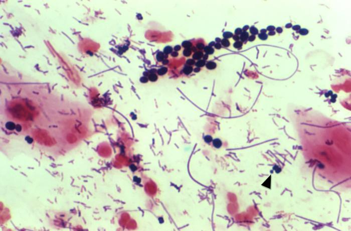
25 Most Commercially Insured Patients. Often several different micro-organisms can be cultured particularly Candida albicans and the Gram-negative bacilli pseudomonas. Candida albicans in Gram-stained wet mounts of vaginal secretions showing both pseudohyphae and budding yeast cells.
Usually your immune system keeps yeast under control. Gardnerella vaginalis is a pleomorphic gram-variable. Although most often caused by a bacterial pathogen it may also result from fungal or viral infection.
Which of the following microorganisms stain well. Alternatively a gram stain of plaques showing large ovoid gram-positive yeast is diagnostic. In many cases it is due to dermatitis of the nail fold.
The most common pathogens detected with a sputum culture are bacteria such as Streptococcus pneumoniae Haemophilus influenzae Staphylococcus aureus and Klebsiella species. Candida albicans is an opportunistic pathogenic yeast that is a common member of the human gut floraIt can also survive outside the human body. It can also be due to the cold sore virus Herpes simplex and the yeast Candida albicans.
Gram-negative bacteria GNB are among the most significant public health problems in the world due to the high resistance to antibiotics. Smears are taken by gently drawing a wooden tongue depressor across the lesion. Glucose fermentation with gas production urea and H 2 S negative lactose positive with exception of strain D - late lactose fermenter.
Source of the culture Gram stain results organism likelihood that the culture was contaminated based on the organisms that are isolated number of organisms that grow and patient gender. Commercial Cash Paying Patients. Produced by gram negative microorganisms Can cause fever.
Fungi are slow-growing eukaryotic organisms that can grow on living or nonliving organisms and are subdivided into molds and yeasts. A centrifuged specimen should be viewed with the aid of Gram stain. Lastly pseudohyphae with a potassium hydroxide stain can be visible.
It is a fungus that lives almost everywhere including in your body. Oral Candidiasis or Oral trush Candida albicans yeast infection on the human tongue close up. Germ tube test.
A second skin biopsy is usually sent for tissue culture for bacteria fungi yeasts and mycobacteria. Candida albicans can be identified presumptively by a simple germ tube test. Specificities of 92 to 98 compared with Gram stain26-28 Vaginal culture and Papanicolaou Pap.
Yeasts can be presumptively identified as Candida albicans when there is the appearance of feet on blood agar in 48 h andor the germ tube is positive. In sections stained with Gram stain gram-negative rods are numerous surrounding necrotic vessels. Candida albicans can have other Candida species White thick cheesy or.
Pay as little as. Actinomyces israelii an anaerobic filamentous gram-positive bacteria is the most common pathogen. Produces white pasty colonies on SBA with feet that look like spider webs.
Depend on a number of factors including. The appropriate selection of e mpiric antibiotic therapy for positive blood cultures is a complex and difficult decision. This is a vaginal yeast infection caused by candida albicans.
Common side effect when using antibiotics or another medicaments. Smears taken from clinical lesions are examined using potassium hydroxide KOH PAS or Grams stain. Join Our Patient Savings Program.
Candida is a commensal organism in the oral cavity. While patients with positive blood cultures may be bacteremic signifying a true. N gonorrhoeae Candida albicans and Herpes simplex virus should be ruled out.
Gram stain of Candida albicans from a vaginal swab. Yeast infections affect different parts of the body in different ways. Vaginitis is defined as any condition with symptoms of abnormal vaginal discharge odor irritation itching or burning.
Smells yeasty on blood agar like bread baking or like beer. Candidiasis is diagnosed by its clinical appearance and by detection of organisms on smears. The small oval chlamydospores are 24 µm in diameter.
May cause just about any type of. Pseudomonas aeruginosa Pseudomonas aeruginosa is a Gram-negative rod-shaped motile organism polar flagella which characteristically produce water-soluble pigments which diffuse through the mediumThe best known are pyocyanin blue-green pyoverdine yellow-green fluorescent and pyorubin red-brown produced by a small proportion of strains. It is detected in the gastrointestinal tract and mouth in 4060 of healthy adults.
In urine gram stain fine with microscope. It is commonly found free living in moist. 298 of the female population was diagnosed with BV with a Gram stain in vaginal fluid.
Escherichia coli Legionella pneumophila Treponema Chlamydia. Bacteria are only partially identified on Gram stain and speciation and susceptibility results will take an additional 24-48 hours after a culture is reported as positive. Sensitivity tests are done on any isolated organisms.

View Image
Clinical Pathology
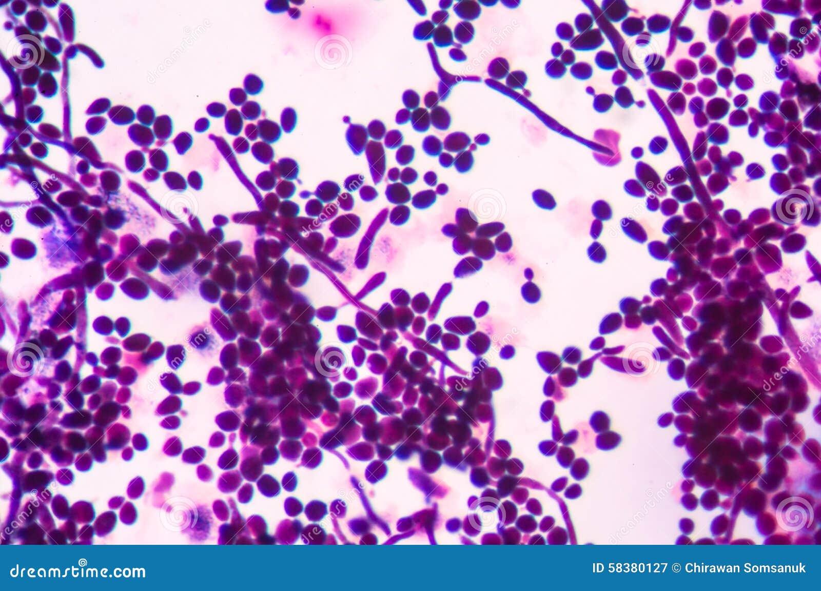
Budding Yeast Cells With Pseudohyphae Stock Image Image Of Yeast Fungi 58380127

Pseudohyphae Yeast Cell Images Stock Photos Vectors Shutterstock
Antifungal Resistant In Non Albicans Candida Species Are Emerging As A Threat To Antenatal Women With Vulvovaginal Candidiasis Biomedical And Pharmacology Journal

Karger Com
Candida Albicans Gram Stain Yeast Stage

Candida Albicans Pathogenesis Diseases Lab Diagnosis Microbe Online


